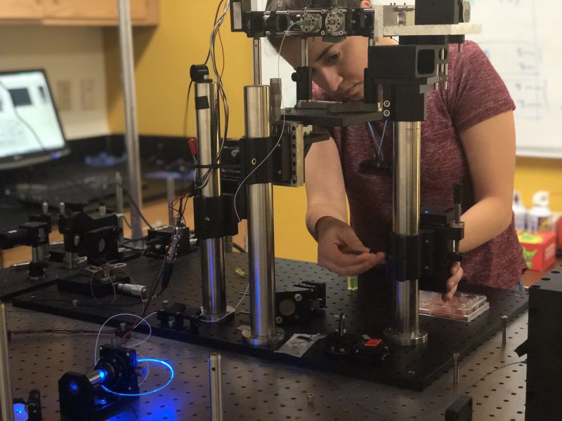
Biomedical engineering doctoral student Zahra Nafar has always had a drive to see the world. While following her passion for traveling, she discovered another love, ophthalmology.
“I basically travel to see different natural scenes. And, I love painting. I mostly paint landscapes and nature views, which are sometimes from the pictures I took during trips,” Nafar says. “I want to see everything, which makes me so interested in how the eye works, how we see and what we see.”
Born and raised in Iran, where she lived comfortably at home with her family, Nafar felt a strong calling to explore the world and document what she saw through photography and paintings. After earning her bachelor’s degree in electrical engineering from the University of Tehran, she set her sights on the United States.
After connecting with Shuliang Jiao, a professor in FIU’s College of Engineering & Computing’s Department of Biomedical Engineering, she was recruited to work as a graduate assistant and work with Jiao in his eye imaging lab.
“The main reason I recruited her was because…she had the passion for the research,” Jiao says.
Now in the United States for the first time, Nafar had to learn how to be truly independent and deal with the challenges of being alone in a foreign country. She would have to overcome language barriers and culture shock.
“Starting over is exciting and scary, but a good scary,” Nafar says. “I have learned a lot just by moving. I have really grown more as a person.”
Nafar, under the direction of Jiao, is currently working on an imaging device called the simultaneous visible-light optical coherence tomography and a fundus autofluorescence imaging system that is patented under Jiao. Nafar is one of Jiao’s first students to generate the preliminary data for grant applications, which has netted $2 million dollars for the research.
Researchers are monitoring early diagnosis and the progression of eye diseases, including age-related macular degeneration, which affects more than 10 million Americans and is the leading cause of vision loss, according to the American Macular Degeneration Foundation. This disease causes the retina to detach leading to people seeing a black circle which spreads across their vision.
The imaging device serves as a diagnostics tool to monitor the progression of macular degeneration. It works by measuring the amount of lipofuscin in the retina, which is an indicator of the disease. This device measures simultaneously with one light source and images the retina and a reference target that consists of lipofuscin as well to compare the measurements to monitor the progression of the disease.
 “I love the fact that I am working hands-on with experimental research, and I am actually helping to build this device so that is what motivates me,” says Nafar.

Meanwhile, Nafar still finds time to pursue her passion for traveling. She goes often on road trips to different cities in various states and visits national parks alongside her husband, whom she met at FIU. She documents it all in her photography, which she posts about on her Instagram page, tripplanningideas. The impression of all her travels can also be seen in her artwork.
When Nafar earns her doctoral degree, she is considering moving with her husband to Europe to pursue a career in ophthalmology or biomedical engineering. She wants to continue seeing the world and all it has to offer while leaving her own contributions to ophthalmology so others can possibly have the chance to see it too.
FIU News, written by: Baely Almonte
